Published on September 17, 2020 By Dr. S.Anoop (Chief Technical Officer and Head - Non Rodent)
Cannulation of the external jugular vein or the central venous line (CVL) access is essential in the laboratory swine models, for pulmonary artery catheterisation and central venous pressure monitoring.
The CVL placement in Laboratory Animals is often performed by surgical cut-down procedures therefore prolonged general anaesthesia is required, moreover surgical cut down at the neck requires significant postoperative management including pain. This method may often lead to permanent closure of vessels and the same vessel cannot be used for subsequent follow up procedure, thus new animals have to be designated for the same procedures.
We at MRIDA (division of Palamur Biosciences) are incorporating MINIMAL INVASIVE procedures following CPCSEA guidelines and encourage 3R Principles while conducting laboratory animal research.
Minimally Invasive vascular catheterisation (1-3) by Needle Puncture is a Refinement as compared to the surgical cut down method and it ensure valid comparisons among animal subjects for human research and it also facilitates the Reduction of the animal numbers for follow up end points as the same animals can be used for the same procedure due to less disruption of architecture of the blood vessels as compared surgical cut down method and reduces animal variation to produce robust data.
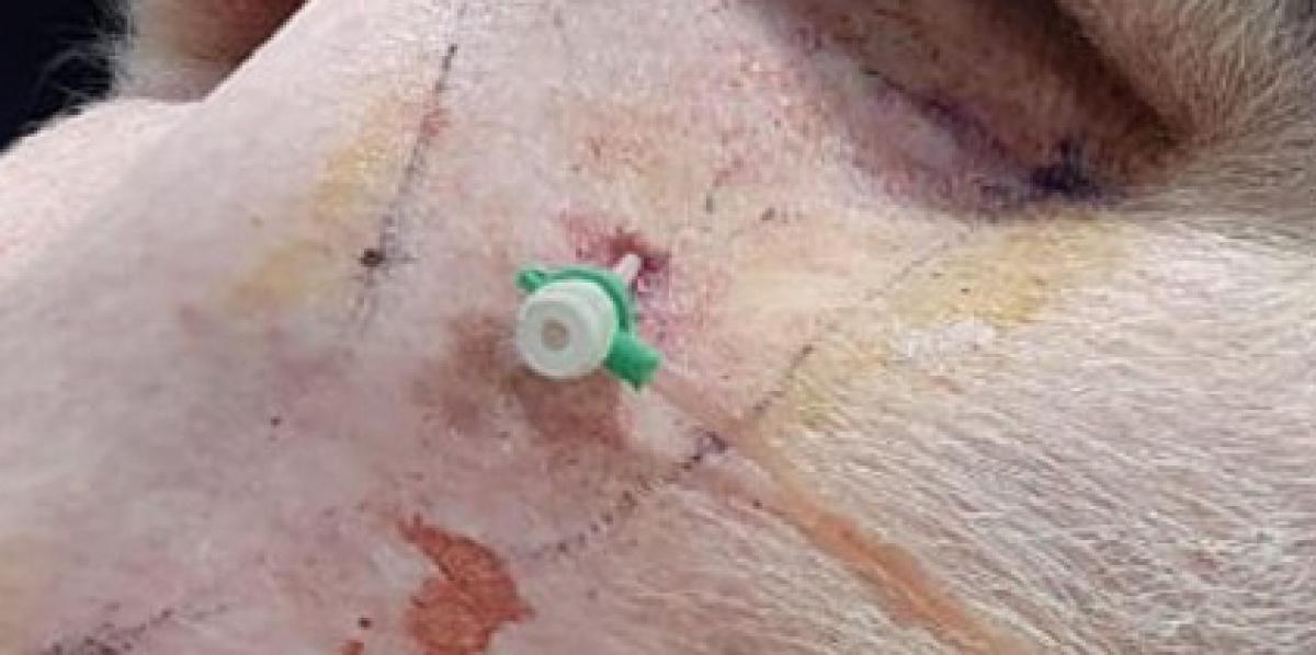
We adopted a TRIANGULATION METHOD for inserting CVL catheters into the jugular vein (JV) using visual and palpable landmarks (4). In this method the mid-point of the triangle is used as guide-point for inserting an 18 G needle into jugular vein and through this needle a guide-wire was inserted into the jugular vein and OTW (over the wire) sheath is inserted.
We confirmed the sheath and catheter placement by intravascular radiography. We often use this technique for central venous pressure (CVP), right ventricle pressure (VP) and pulmonary pressure (PV) during procedures of cardiovascular and cardiac devices implantation
- Video-1: Jugular Vein to right ventricle and pulmonary arteries contrast angiography
- Video-2: Catheter placed in right ventricle for right ventricle pressure
- Video 3: Catheter placed in pulmonary artery for pulmonary artery pressure
References:
- Carroll JA, Daniel JA, Keisler DH, Matteri RL. Non-surgical catheterisation of the jugular vein in young pigs. Lab Anim 1998;33:129 – 34
- Matte JJ. A rapid and non-surgical procedure for jugular catheterisation of pigs. Lab Anim 1999;33:258–64
- Zanella AJ, Mendl MT. A fast and simple technique for jugular catheterisation in adult sows. Lab Anim 1992;26:211–13
- Flourney WS, Mani S. Percutaneous external jugular vein catheterisation in piglets using a triangulation technique Lab Anim 2009 Oct;43(4):344-9


