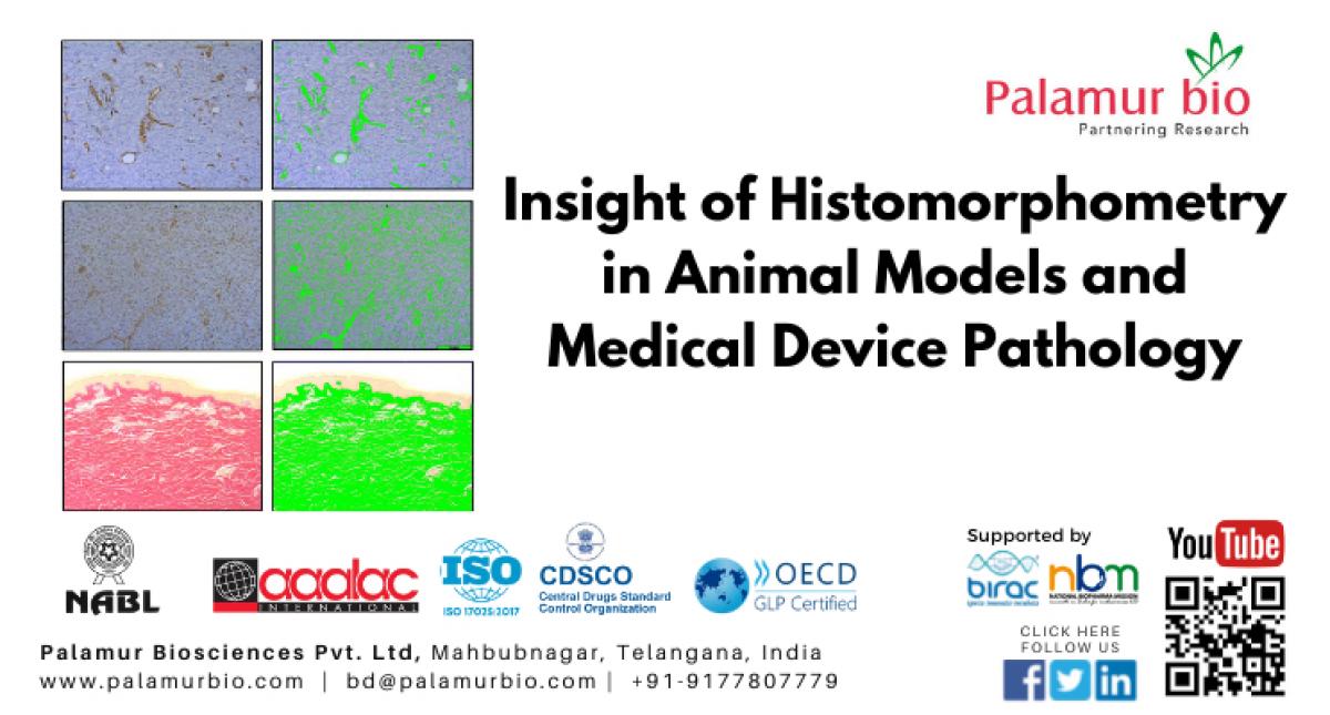
The main aim of any histopathological investigation is the identification of a pathological process; therefore special diagnostic features are necessary. Histomorphometry is broadly defined as the measurement of the shape or form of a tissue. Histomorphometry is the method used to quantify histological features tissues. It is an important tool in animal model studies like IPF (Idiopathic pulmonary fibrosis), NASH (Nonalcoholic Steatohepatitis), PAH (Pulmonary arterial hypertension), Liver fibrosis, Colitis, EAE (Experimental autoimmune encephalomyelitis), Psoriasis, Arthritis, neoplasm and others.
It is also very useful in analysis of medical device pathology like coronary and femoral stents, muscle and bone implants, tissue and mechanical heart valves etc. It is useful tool in semi-quantification of immune staining (Immunohistochemistry). Semiquantitative scoring systems are widely used to convert subjective perception of IHC-marker expression by pathologists into quantitative data, which is then used for statistical analyses and establishing of the conclusions.
Histomorphometric examination of tissues is based on quantitative measurements of microscopic organization and structure which used to provide information on cellular responses (e.g., Migration, Inflammation), tissue pathology (Fibrosis, Lipidosis, atherosclerotic lesions, tumor growth) as well as metabolic bone disturbances.
Histomorphometry measurement is important in post stent histopathology to study the in-stent restenosis as a result of neointimal hyperplasia.
The long-term clinical efficacy of intracoronary stenting is limited by restenosis, which occurs in 15% to 30% of patients (1). Stent induced mechanical arterial injury and a foreign body response to the prosthesis incite acute and chronic inflammation in the vessel wall, with elaboration of cytokines and growth factors that induce multiple signaling pathways to activate smooth muscle cell (SMC) migration, proliferation and accumulation of extracellular matrix.
Drug-eluting stents (DES) dramatically decreased in-stent restenosis via inhibition of SMC proliferation. In the pig model, Carter et al. (1994) demonstrated that the inhibition of neointimal hyperplasia was obtained at one month by sirolimus-eluting stents. However, at 6 months, the proliferation rate was significantly higher in arteries with sirolimus-eluting stents than controls, suggesting late restenosis.
MRIDA is a division in Palamur Biosciences Pvt Ltd., and a reference institute for medical device studies. After completion of our implant is subjected to histopathology and histomorphometry. Department of Pathology is well equipped with advanced instrument for standalone pathology services.
Histomorphometry or image analysis is an important tool in the assessment of many types of histopathology changes. Digital photomicrographs are captured and imported in to image analysis software. For histomorphometry analysis, sections were stained with hematoxylin and eosin or special stain or IHC. The semi-quantitative scoring is performed for inflammation, vascular injury, fibrin deposition and, loss of smooth muscle and endothelium. Histo-Morphometrically medial area, neointimal area and percent stenosis calculated. Steps involved in histomorphometry as follows:
- Mehran R, Dangas G, Mintz GS, et al. Patterns of in-stent restenosis: angiographic classification and implications for long-term clinical outcome. Circulation. 1999;100:1872–1878.
- 2. Carter AJ, Laird JR, Farb A, et al. Morphologic characteristics of lesion formation and the time course of smooth muscle cell proliferation in a porcine proliferative restenosis model. J Am Coll Cardiol. 1994; 24:1398–1405.


