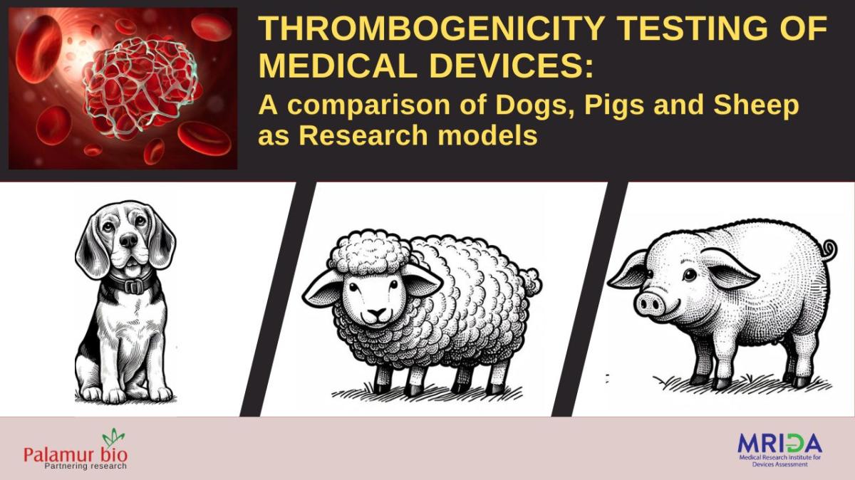
Authors: Dr. D.C. Sharma & Dr. Sachin Bansal
ABSTRACT:
Thrombogenicity is the tendency of a material or device to induce blood clotting, which can lead to serious complications such as device failure, thromboembolism, stroke, or myocardial infarction. Thrombogenicity testing is an essential part of the preclinical evaluation of medical devices that come in contact with blood, such as stents, catheters, valves, grafts, and ventricular assist devices. Thrombogenicity testing aims to assess the potential for material-mediated or geometry-mediated thrombus formation on the device surface or downstream from the device. Thrombus formation can result from various factors such as activation of platelets, coagulation factors, complement system, leukocytes, endothelial cells, and inflammatory mediators. Thrombogenicity testing also evaluates the effects of anticoagulant or antiplatelet therapy on device performance and safety. In this blog, we review the current methods and challenges of thrombogenicity testing using large animal models (dogs, pigs, and sheep), which are considered more relevant and predictive than small animal models (such as rodents) for human applications. We also discuss the regulatory requirements and standards for thrombogenicity testing of medical devices in the US and internationally.
HIGHLIGHTS:
- Thrombogenicity testing of medical devices is performed to assess the potential for blood clot formation and adverse events in patients.
- Large animal models (dogs, pigs, and sheep) are preferred over small animal models for thrombogenicity testing because they have more similar hemodynamics, hemostasis, and anatomy to humans.
- Thrombogenicity testing methods include in vitro assays, ex vivo perfusion systems, and in vivo implantation studies. Each method has its advantages and limitations in terms of cost, complexity, reproducibility, and clinical relevance.
- Regulatory agencies such as the US Food and Drug Administration (FDA) and the International Organization for Standardization (ISO) provide guidance and standards for thrombogenicity testing of medical devices. The ISO 10993-4 standard specifies the general requirements for evaluating the interactions of medical devices with blood, while the FDA provides device-specific recommendations and expectations for thrombogenicity testing.
- The requirements of different large animal models for thrombogenicity testing depend on various factors such as device type, target site, implantation duration, and anticoagulation regimen. Dogs are commonly used for cardiovascular devices but have high variability in thrombus formation (particularly for the small size devices such as CVL lines compatible to the vasculature of the dogs). Sheep are suitable for long-term implantation studies as well as for implantation of large devices as sheep's vasculature is bigger in size than that of dogs, but require careful anticoagulation monitoring because of high vulnerability of RBCs to get clotted. Pigs are widely used for all type of implantations (active as well as cardiac implants and conduits for the vascular implantation). They are specifically more compatible to the coronary interventional devices.
THROMBOGENITY TESTING METHODS:
Thrombogenicity testing methods include in vitro assays, ex vivo perfusion systems, and in vivo implantation studies. Each method has its advantages and limitations in terms of cost, complexity, reproducibility, and clinical relevance.
In vitro assays are performed using blood samples or isolated blood components (such as platelets or plasma) that are exposed to device materials or extracts under static or dynamic conditions. They can measure various parameters such as platelet adhesion and activation, coagulation activation and inhibition, fibrin formation and degradation, complement activation, leukocyte adhesion and activation, and cytokine release. In vitro assays are relatively simple, fast, and inexpensive to perform. They can also be used to screen different materials or device designs before animal testing. However, these have limited ability to mimic the complex interactions between blood, device, and vessel wall in vivo. They also do not account for the effects of systemic factors such as inflammation, infection, or medication on thrombogenicity.
Ex vivo perfusion systems are performed using whole blood or plasma that is circulated through a closed-loop system containing a device or a segment of a vessel with a device implanted. They can simulate more realistic hemodynamic conditions than in vitro assays by controlling parameters such as flow rate, pressure, temperature, and oxygenation. They can also measure parameters such as thrombus formation and embolisation, blood cell damage, and device patency and function. These systems are more complex, time-consuming, and expensive than in vitro assays. They also require fresh blood or plasma from animals or humans, which can introduce variability and ethical issues.
In vivo implantation studies are performed using large animals that receive a device implantation in a target vessel or organ. In vivo implantation studies can evaluate the thrombogenicity of a device in the most relevant physiological and pathological context. These studies are the most complex, time-consuming, and expensive methods of thrombogenicity testing. They also require ethical approval and animal welfare considerations.
LARGE ANIMAL MODELS FOR THROMBOGENICITY TESTING:
- Dogs are commonly used as a large animal model for thrombogenicity testing of cardiovascular devices, such as stents, catheters, valves, and grafts. Dogs have similar hemodynamics, coagulation cascade, and platelet function to humans. However, dogs also have limitations, foremost is the size of the device under test such as the TAVR balloons and Valvuloplasty balloons that are available in big sizes according to the human use but are unable to get evaluated using dogs due to smaller size vasculature limitations. In addition, dogs have high variability in thrombus formation, and low sensitivity to detect intermediate thrombotic materials. Therefore, the FDA recommends using relevant animal models that reflect the intended clinical use and patient population of the device.
- Sheep are another large animal model for thrombogenicity testing of cardiovascular devices, especially those that require long-term implantation or integration with the vessel wall. Sheep have comparable vessel diameter, blood pressure, and blood flow rate to humans. However, sheep also have some challenges, such as difficulty in achieving appropriate anticoagulation, high susceptibility to infection and inflammation, and species-specific thrombotic events, and being a ruminant species challenging to perform implantation procedures under anaesthesia. Therefore, the FDA recommends careful monitoring of anticoagulant therapy and descriptive narratives from the pathologist or veterinary pathologist to understand the causes of thrombosis.
- Pigs are a widely used large animal model for thrombogenicity testing of cardiovascular devices, especially those that involve coronary intervention or stenting. Pigs have similar hemodynamics, anatomy, and biologic response to humans. Pigs can also accommodate larger or multiple devices than dogs or sheep. The FDA recommends using domestic swine over miniature swine for the size compatibility issues.
REGULATORY REQUIREMENTS AND STANDARDS FOR THROMBOGENICITY TESTING:
IN-VIVO THROMBOGENICITY TESTING MODELS AT PALAMUR BIOSCIENCES:
EXPERIMENTAL DESIGN AND END POINTS FOR IN-VIVO THROMBOGENICITY TESTING:
- The animal is prepared for the thrombogenicity testing of implantable device under proper analgesia and gaseous anaesthesia.
- Percutaneous approach using Seldinger method/vessel exposure is used to insert sheath of 14F into femoral artery, if it is difficult to use percutaneous approach a cut down at the groin to expose femoral artery is be done followed by 14F sheath insertion.
- ACT measurement is be done pre and post heparinisation (The heparinisation to be done to keep ACT values between 250 to 550 seconds). The first bolus dose is be given at 80-120 IU/kg IV/IA and subsequent doses will be titrated based on ACT values.
- The aplitzer 0.035-inch diameter and 260cm length guidewire is used to navigate the test item into the ascending aorta and left ventricle.
- The test item is navigated OTW to the target site, inflated and deflated and kept deflated for at the most 30 sec (time duration can be reduced based on ECG complications) then deflated and removed for thrombogenicity evaluation.
- In accordance with clinical interventional technique, the sheaths will be flushed with heparinized saline every 20 min.
- During this time the ACT is checked approximately every 20 minutes to ensure clinically relevant ACT levels are maintained.
- The radiographic cine image of the Valvuloplasty balloon is taken to confirm the placement in the thoracic ascending aorta.
- At the conclusion of the simulated use, withdrawal, and thrombus evaluation on withdrawn the Valvuloplasty balloon will be performed with photography for assessment of thrombogenicity on the device.
- The animal is humanly euthanised to harvest the target site vessels and the navigation vessels for any evidence of the thrombogenic activity of the test device.
- Post euthanasia animals is subjected to a comprehensive necropsy, defined as gross external and internal organs examination for the limited organs pertaining to the ISO guideline. The target sites such as ascending aorta in the current protocol is given more thorough examination for any lesions of thrombogenic importance.
- Lesions are documented and collected wherever observed, immersion fixed in NBF pending processing for histology.
- Tissues without macroscopic findings may also be sampled to assist in the evaluation of animal health.
- Images of target site (ascending aorta) are taken in order to show the presence/ absence of acute thrombosis or tissue injury.
- In addition to the above important evaluations pertaining to the thrombogenicity, clinical evaluation of D Dimer along with pre-and-post deployment haematology and clinical biochemistry is performed on the blood samples collected time to time during the procedure.
- The photographic evidence of the withdrawn test article is taken to score the thrombus formation as per ISO 10993-4.
- The conclusive statement is structured by the study director based on the clinical evaluation, in-life photographic thrombus observation score on the withdrawn test device and necropsy/histopathological evaluation of the harvested target site vessel.
- Scoring of thrombus formation due to device deployment as per scoring schema A of ISO 10993-4:2017.
Image of the Thrombus Formation and Scoring:


